Introduction
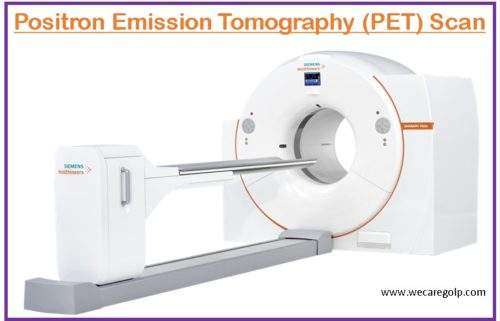
PET scan (positron emission tomography Scan) is a functional scanning technique that uses radiotracers (radioactive substances), a special camera, and a computer to project and measure variations in metabolic processes, and other physical activities inside the body including blood flow, local chemical composition, as well as absorption. The scan reveals both normal and abnormal metabolic or biochemical activities of the tissues and organs earlier than other imaging tests like CT scan or MRI. The PET images are also integrated with CT or MRI and are called PET-CT or PET-MRI scans.
- Before the PET scan, an injection of a little amount of a radioactive tracer or glucose called fluorodeoxyglucose-18 (FGD-18) is given to the patient.
- Cancerous cells absorb more radiotracer glucose than healthy cells which is known as hot spots.
- The scan then takes x-rays of the body from different angles and the computer merges the PET and CT images to produce a detailed three-dimensional (3D) result showing abnormalities like tumors.
- PET scans also help in determining the functionality and strength of the internal body organs.
Purposes of PET Scan
- To detect malignancies and determine whether the tumor has spread in the body.
- To evaluate the effectiveness of treatment and determine the prognosis.
- To determine if cancer has reemerged after treatment or not.
- To assess the metabolism and viability of tissues.
- To determine the effects of myocardial infarction or a heart attack on the heart.
- To identify muscle areas of the heart that would benefit from angioplasty or coronary artery bypass surgery (when a PET scan is done with a myocardial perfusion scan).
- To evaluate abnormalities in the brain like tumors, seizures, memory disorders, and other central nervous systems (CNS) disorders.
Indications of PET Scan
- Cancer (lungs cancer, breast cancer, Hodgkin’s lymphoma, non-Hodgkin’s lymphoma, cervical cancer, thyroid cancer)
- Brain and mental health disorders (Alzheimer’s disease, brain tumors, Parkinson’s disease, epilepsy, stroke, brain injuries, dementia, anxiety, schizophrenia, depression)
- Heart diseases (heart attack, coronary artery disease, other heart diseases)
- Infections (vasculitis, osteomyelitis, diabetic foot syndrome, peritoneal tuberculosis)
Procedure of PET Scan
Before the Procedure
- Inform the doctor if the patient is taking any prescribed, over-the-counter (OTC), or supplemental medications.
- Avoid strenuous physical activity, such as exercise, and deep-tissue massages in the 24 to 48 hours before the test.
- Diet: Patients must take a low carbohydrate and zero sugar diet 24 hours before the procedure.
- Not allowed foods and beverages are cereal, pasta, bread, rice, milk and yogurt, fruit and fruit juices, alcohol, caffeinated beverages, candies, chewing gum, and mints.
- Allowed foods are usually non-starchy like meat, tofu, nuts, and vegetables like carrots, asparagus, broccoli, salad greens, squash, etc.
- Patients are not allowed to eat or drink anything 6 hours before the PET scan whether sedatives are given during the procedure or not. They can take sips of water and medicines as prescribed.
- Inform the doctor if the patient is pregnant or breastfeeding because the test may be unsafe for the fetus and they cannot breastfeed for 24 hours after the test.
- If patients have diabetes, they should take the normal dose of insulin and a light meal 4 hours before the schedule of a scan.
- Patients need to wear hospital gowns.
- Any pieces of jewelry including body-piercing are to be removed because metal can interfere with the scanner but medical devices like pacemakers and artificial hips will not affect the test results.
During the Procedure
- Before the scan, tracers are given through intravenous, oral, or inhalation routes. It will take about an hour (depending on the area of the body being scanned) to absorb the tracers.
- The patient should relax, limit any movement, and try to stay warm and avoid television, music, and reading before the brain scan.
- Patients are advised to lie down on the narrow table attached to a PET machine which is slid into the scanner. The patient should stay still and hold their breath several times during the scan.
- The procedure usually completes in 30 to 45 minutes but if the patient is having multiple tests, this may take up to about 3 hours. The patient can hear buzzing and clicking noises during the test.
After the Procedure
- The patient can continue their normal activities after the scan including driving.
- They should have plenty of water to wash the radioactive substance/dye out of the body.
Limitations of PET Scan
Even though the amount of radiation given during a standard PET scan is considered safe, minimal radiation exposure also has the potential risk of tissue damage that could give rise to malignancy in later life.
- The image resolution of a sole PET scan may not be as high as that of an MRI or CT scan.
- The CT component of a PET-CT scan also adds to the amount of radiation.
- The procedure is time-consuming because it can take many hours to days for the radiotracer to concentrate on the area that needs to be scanned. The imaging procedure may also take several hours to be completed.
- The radiotracer quickly becomes less radioactive progressively and usually be washed off naturally from the body within a few hours, so the procedure must be done at the exact time.
- As patients are radioactive for certain hours after a PET scan, they are encouraged not to come in close contact with pregnant ladies, small kids, and infants.
- Altered blood sugar or blood insulin levels in patients with diabetes may adversely affect the test results.
- A very obese patient may not fit properly into the conventional PET Scanner.
Complications of PET Scan
- Anaphylaxis
- Contrast-induced nephropathy
- Hyperglycemia (in the case of diabetics)
Interpretation
- On PET scans, cancer cells are seen as large, bright spots with a dark color due to a higher metabolic rate than normal cells.
Summary
- PET scan utilizes high-frequency radiation to generate a 3D image of the internal parts of the body.
- Specialized radioactive contrast dyes are used in the scanning process to detect the growth or abnormalities of a micro-organism at the cellular level.
- The PET scan is most used to identify and treat malignancies at an early stage than CT scans and ultrasounds.
- The PET scan is usually combined with MRI and CT scan. Although it has minimal radiation exposure, it is considered a safe procedure.
References
- Kapoor, M. (2022). PET Scanning. Statpearls. https://www.statpearls.com/ArticleLibrary/viewarticle/27066
- Unterrainer, M., Eze, C., Ilhan, H. et al. (2020). Recent advances of PET imaging in clinical radiation oncology. Radiation Oncology, 15, 88. https://doi.org/10.1186/s13014-020-01519-1
- Slough, C., Masters, S. C., Hurley, R. A., & Taber, K. H. (2016). Clinical positron emission tomography (PET) neuroimaging: advantages and limitations as a diagnostic tool. The Journal of neuropsychiatry and clinical neurosciences, 28(2), A4, 67-71. DOI: 10.1176/appi.neuropsych.16030044
- Cleveland Clinic. (2022, Oct 19). PET Scan. Retrieved on 2023, Mar 16 from https://my.clevelandclinic.org/health/diagnostics/10123-pet-scan
- Krans, B. (2021, Dec 16). What Is a Positron Emission Tomography (PET) Scan? Healthline. Retrieved on 2023, Mar 16 from https://www.healthline.com/health/pet-scan

