Introduction
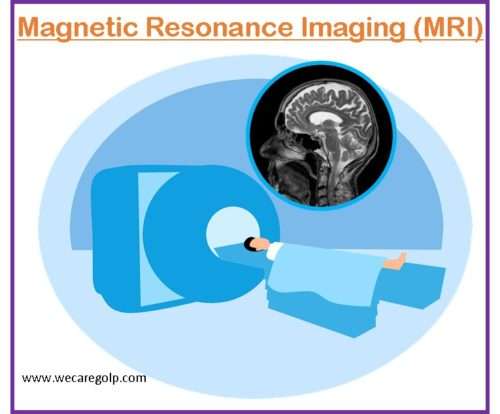
Magnetic resonance imaging (MRI) scan is a type of scan that uses a large magnet or strong magnetic field, radio waves, and a computer to create a thorough, cross-sectional image of internal body organs, tissues, and different structures.
- An MRI scan is done for the assessment of any part of the body like the
- Brain and spinal cord,
- Bones and joints,
- Heart and blood vessels,
- Breasts, and
- Other internal organs, such as the liver, uterus, prostate gland, etc.
- MRI scans can be useful in
- Diagnosing health problems,
- Planning treatments, and
- Assessing the effectiveness of treatment accordingly.
- MRI does not have ionizing radiation and is capable of very precise tissue differentiation.
- MRI differs from computed tomography (CT), as CT uses X- rays.
Mechanism of MRI Scan
- The MRI machine is a large and cylindrical machine that produces a powerful magnetic field throughout the person and pulses of radio waves are sent by a scanner.
- The radio waves strike the nuclei of the protons in our body out of their natural/normal position and align in an axis.
- As the nuclei rearrange into proper position, they transmit radio signals which are accepted by a computer that interprets and transforms them to create a two-dimensional (2D) image of the body part being scanned.
- These images are then seen on the computer screen.
- Conventional MRI machines have narrow tunnels, but new ones are more spacious.
- MRI scans can take 30 to 120 minutes.
Types of Magnetic Resonance Imaging
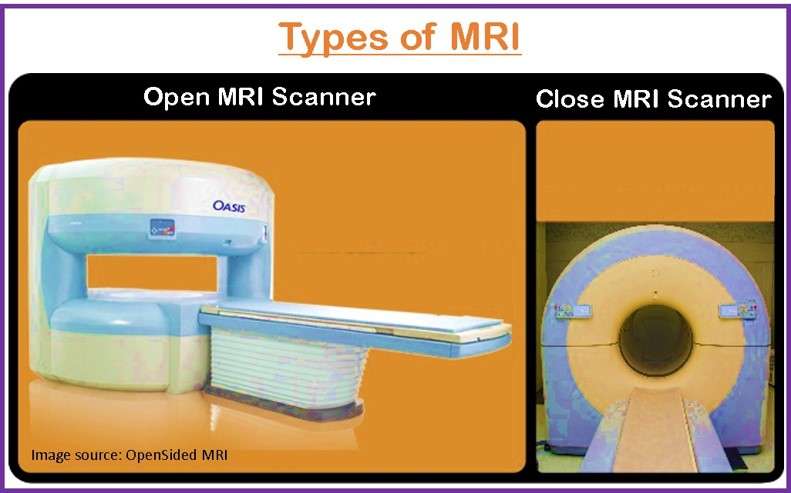
- A closed or regular MRI scanner has a large cylindrical tube encircled by a magnet in which the person is kept on the sliding table to slide into the tube.
- In an open MRI machine, the patient lies between two flat magnets that are positioned above and under the patient. Open MRI is a more convenient type of scanner for claustrophobic (fear of confined or enclosed spaces) or obese persons, but the image quality is better in the closed type.
Indications of Magnetic Resonance Imaging
MRI has varieties of indications and it is also a suitable method for the staging of most cancers. Some of the indications for MRI are as follows:
- Nervous system
- Posterior fossa pathology
- Cerebrovascular diseases
- Demyelinating diseases e.g., multiple sclerosis
- Dementia
- Infectious disease
- Brain tumors and aneurysms
- Stroke
- Seizure disorders, etc.
- Cardiovascular system
- Myocardial ischemia
- Cardiomyopathies
- Carditis
- Congenital heart diseases
- Vascular diseases
- Angiography of blood vessels stenosis, Aneurysms, malformations, etc.
- Musculoskeletal system
- Spinal imaging
- Knee and shoulder injuries
- Diseases of joints
- Tumors of soft tissues, etc.
- Lesions of the liver, pancreas, bile ducts, and other GI system
- Iron overload
- Breast cancer
Additional magnetic resonance technology
- Magnetic resonance angiography (MRA) is done to analyze blood flow through the arteries that detect intracranial aneurysms and vascular malformations within the brain, spinal cord, or other parts of the body.
- Magnetic resonance spectroscopy (MRS) is done to detect chemical abnormalities in body tissues such as stroke, head injury, Human Immunodeficiency Virus infection of the brain, tumors, multiple sclerosis, Alzheimer’s disease, etc.
- Functional magnetic resonance imaging of the brain (fMRI) is done to determine the specific location of the brain where a certain function occurs like speech or memory. The general areas of the brain in which such type of functions occurs are the same in all human beings but the exact location may differ in each person.
Contraindications of Magnetic Resonance Imaging
MRI is contraindicated in those patients who have:
- Pacemaker or artificial heart valves
- Any type of implantable pumps, such as an insulin pump
- Vessel coils, filters, stents, or clips
- Suspected or confirmed pregnancy
- Body piercing
- Permanent eyeliner or tattoos
- Bullet wound
- Implants, prostheses, dental braces, or wires
- Work with metal (a metal grinder or welder)
- Other metallic fragments anywhere in the body
- Difficulty lying down for 30 to 60 minutes
Procedure
Before the Procedure
- Depending on the area being scanned patients may be kept NPO for 4 hours or they are allowed to eat and drink normally. Sometimes patients are advised to have plenty of fluids.
- The health and medical history of the patient is taken beforehand to ensure safety of the patient.
- The patient should wea r hospital gown or clothes without metal buttons, zips, wire, belts, buckles, fasteners, etc. during the procedure.
- To prevent the effects of strong magnets and radio wave signals, it is very important to ensure that there are no metal objects outside and inside the patient’s body like:
- Watches, jewelry, and hairpins
- Body piercings
- Artificial dentures
- Hearing aids
- Wigs that contain metal traces
- Hairpins and clips
- Medicine patches like nicotine or hormone patches or patch insulin pumps
- Glucose monitors etc.
During the Procedure
- The patient will be placed on a scan table.
- The scanner makes extremely loud tapping noises at certain times during the MRI scan which is due to the electric current being turned on and off in the scanner coils. Thus, earplugs or headphones may be provided for the patients to listen to music to make them comfortable.
- Patients are advised not to move and remain still during the entire scan for clear images.
- If the patient needs contrast, the radiologist/technologist gives it via IV injection before you undergo the MRI.
- If the patient has claustrophobia, he/she may get sedative drugs or anesthesia during the procedure.
After the Procedure
Patients are advised to:
- The patient can get up and move out slowly from the scanner table to prevent lightheadedness or dizziness related to the prolonged flat position.
- If sedatives were given, the patient is suggested to take rest until it has worn off. S/ he also needs someone to drive him/her home.
- Any side effects or reactions to the contrast dye will be observed, such as itching, rashes, edema, or difficulty in breathing if MRI was done with contrast.
- The indication of an infection or allergy like pain, redness, and/or swelling at the intravenous site will be watched.
- The patient can resume usual activities and diet unless contraindicated.
Side Effects of of Magnetic Resonance Imaging
There are no side effects of MRI. In rare cases, some patients may experience the following side effects due to contrast material.
- Headache
- Pain at the injection site
- Nausea
- Allergic reaction
- Thickening and hardening of the skin and organs (nephrogenic systemic fibrosis) in kidney patients
Precautions
MRI is considered a safe imaging procedure but some precautions are required in the MRI scanning room because an MRI magnet has a local magnetic field that is 30,000 times stronger than the magnetic field of the earth.
- Any magnetic object can come close to the scanner and create a safety hazard as well as image artifacts.
- There is also the risk of causing movement, generating heat or a current, or causing malfunction of devices.
- The items may become projectiles or get stuck in the machine and may cause serious injury or the death of a patient who is inside the scanner.
Summary
- MRI is a noninvasive diagnostic imaging procedure that is best for the imaging of soft tissues with high-quality imaging and good anatomic details.
- MRI has strong magnetic fields so that no metals or electronic devices inside or outside the body are allowed into the scan room.
- MRI does not use ionizing radiation so it has no radiation hazards.
- It helps to detect more precise pathologies of soft tissues, blood vessels, and other systems.
- It is considered a safe procedure but is time-consuming and expensive.
References
- Cleveland Clinic. (2022, Sep 05). MRI (Magnetic Resonance Imaging). Retrieved on 2022, March 05 from https://my.clevelandclinic.org/health/diagnostics/4876-magnetic-resonance-imaging-mri
- John Hopkins Medicine. Magnetic Resonance Imaging (MRI) of the Bones, Joints, and Soft Tissues. Retrieved on 2022, March 06 from https://www.hopkinsmedicine.org/health/treatment-tests-and-therapies/mri-of-the-bones-joints-and-soft-tissues
- University of California, San Francisco (UCSF). Prepare for Magnetic Resonance Imaging (MRI). Retrieved on 2022, March 06 from https://radiology.ucsf.edu/patient-care/prepare/mri
- National Institute of Biomedical Imaging and Bioengineering (NIBIB). Magnetic Resonance Imaging (MRI). Retrieved on 2022, March 06 from https://www.nibib.nih.gov/science-education/science-topics/magnetic-resonance-imaging-mri