Introduction
Myelography is an invasive diagnostic procedure used to examine the spinal canal using x-rays. The method was first described by Sicard and Forestier in 1921.
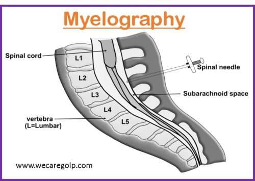
- A contrast dye is injected into the spinal canal through a hollow spinal needle into the subarachnoid space, for a better view and assessment of the
- Nerve roots,
- Spinal cord,
- Spinal nerves, and
- Other soft tissues.
- The resulting imaging is called a myelogram.
- Myelography is particularly useful for showing even subtle cases of nerve root compression using either of these two imaging tests:
- Fluoroscopy: It is a medical procedure that makes a real-time video of the movements inside a part of the body by passing x-rays through the body over a while.
- CT scan (computerized tomography): A CT scan combines a series of X-ray images taken from different angles around the body and uses computer processing to create cross-sectional images (slices) of the soft tissues, blood vessels, and bones.
Indications of Myelography
- Invasive defects of the spinal canal caused by tumors or herniated intervertebral disks into the lower thoracic and lumbar regions
- Tumor, space-occupying lesions, or infection
- Abnormalities of the blood vessels in the spinal cord
- Degenerative diseases of the central nervous system (CNS)
- Infection, inflammation of the meninges as well as soft tissue
- Malformation of the spinal cord
- Syringomyelia (a disorder in which a fluid-filled cyst called a syrinx form within the spinal cord)
- Injury of spinal nerve roots
- Spinal stenosis
- Bone spurs
- Arachnoiditis ( inflammation of a delicate membrane that covers the nerve roots in the lower spine)
- Arthritic disks
Contraindications of Myelography
Absolute contraindications
- Increased intracranial pressure
Relative contraindications
- Grave deformity of the thoracic cage
- Pregnancy
- Uncooperative patient
- Connective tissue disorders (such as Marfan’s and Ehlers-Danlos syndromes)
- Uncontrolled diabetes (ketoacidosis): nausea and vomiting can complicate post-procedure care after myelography.
Procedure
Before the Procedure
- If the patient has diabetes, notify the physician. The patient may take his usual dose of insulin.
- If the patient takes antihypertensive drugs, he may continue it. But, other medications may need approval from the physician.
- The patient cannot drive home after the test so an assistant may be needed.
- Copies of previous relevant X-rays, CT scans, or MRIs for review and comparisons with myelography images should be carried to the imaging center.
- NPO (Nil per Oral) after midnight before the procedure. Clear liquids are usually acceptable.
- Anticoagulants may need to be held before myelography:
- Motrin, aspirin, and nonsteroidal anti-inflammatory drugs: before seven days
- Coumadin: before five to seven days
- Lovenox: before 24 hours
- Heparin: before four hours
- Any psychological drugs such as antidepressants (MAO inhibitors) and anti-nausea medication — before 48 hours and for 24 hours after the test.
- Remove jewelry before the test.
During the Procedure
- The patient should wear a hospital gown and lie on the stomach or side of the examination table.
- Monitors will be placed to measure heart rate, blood pressure, and oxygen level in the blood.
- Intravenous fluids may be given.
- A local anesthetic is injected into the skin of the patient’s back.
- A spinal needle is used to inject the X-ray dye into the spinal column.
- A sample of spinal fluid may be taken for testing.
- The examination table may be tilted in different ways to help distribute the dye in the spine.
- Multiple X-rays are taken.
- A computed tomography (CT) scan or magnetic resonance image (MRI) or both may be done after the test.
After the Procedure
- The patient will be observed for one hour immediately following the myelogram.
- Any of the following changes should be notified to the neuro-radiologist immediately.
- Numbness and tingling of the legs,
- Blood or other drainages with/or pain at the injection site,
- Headaches,
- Nausea or vomiting,
- Inability to urinate,
- Fever (above 100.4 degrees F),
- Stiff neck, etc.
- If headaches persist for more than 24 hours after the procedure or worsen during position it should also be informed.
- After myelography, patients are advised:
- To take rest lying down for 4 hours.
- No strenuous activities for 24 hours, and avoid bending movements for 48 hours.
- Drink plenty of fluids to clear the contrast from the body.
- Breastfeeding patients should not feed their breast milk to the child for a day after the test.
- In general, anticoagulant drugs may be started the day following the procedure.
Benefits of Myelography
- Myelography is commonly done with a CT scan in cases where the spinal cord, subarachnoid space, nerves, discs, and other soft tissues need to be examined in detail that cannot be identified by MRI or standard X-ray.
Risks of Myelography
- Infection
- Cerebrospinal fluid (CSF) leakage
- Bleeding in the spinal canal
- Spinal headache
- Nausea, vomiting, and dizziness
- Allergic reaction to the contrast dye
- A pregnant woman may have associated risks of radiation exposure.
- Risk of contrast medium when part of the bevel of the injected needle is in the subarachnoid space and another part is in the subdural space. The contrast medium would flow freely in the subdural space exactly as same as in the subarachnoid space.
Summary
- Myelography is a form of imaging test performed to evaluate the subarachnoid space, spinal cord, and nerve roots within the spinal canal.
- It is useful for assessing the spine after surgery and for disc abnormalities in patients who cannot undergo MRI.
- Some relative contraindications for a myelogram are pregnancy, uncooperative patient, connective tissue disorder, etc.
- Myelograms have some side effects like headache, CSF leak, exacerbation of back and leg pain, infection, etc.
- Specific post-procedural care includes bed rest for 24–48 hours and plenty of fluid.
References
- Patel D. M., Weinberg, B. D., Hoch, J. M. (2020, Feb 14). CT Myelography: Clinical Indications and Imaging Findings. RadioGraphic, 40(2). https://doi.org/10.1148/rg.2020190135
- Gilcrease-Garcia, B., El-Feky, M., Murphy, A., et al. Myelography. (2023, Feb 2). Myelography. Radiopedia.org. https://doi.org/10.53347/rID-61548
- MedlinePlus. (2021, Nov 12). Myelography. Retrieved 2023, Feb 2 from https://medlineplus.gov/lab tests/myelography/
- Sharma, M. et. al., (2020). Comprehensive Textbook of Medical-Surgical Nursing. Samikshya Publication, Kathmandu
- Mandal, G.N. (2012), Textbook of Adult Nursing. Makalu Publication House. Kathmandu