Introduction
An electroencephalogram (EEG) is a diagnostic test which measures the electrical activity of the brain or the electrical signals sent between brain cells. This electrical activity is seen on a computer screen as ‘brain waves’, which are interpreted by a specialist doctor or a technician. EEG is usually done to identify seizures in clinical settings but can also be used by researchers to explore various brain functions and their relation to human cognition and behavior. The first human EEG was recorded by Hans Berger (German physiologist and psychiatrist) in 1924.
Indications
- EEGs is performed to diagnose seizures associated with:
- Seizure disorders (e.g., epilepsy)
- Dementia
- Narcolepsy (sleep disorders)
- Encephalitis
- Stroke
- Brain tumor, inflammation, and infection
- Brain injury
- Encephalopathy
- It is also done to:
- Determine the best treatment option based on the types of seizures
- Identify and locate a suspected brain lesion
- Evaluate periods of unconsciousness
- Predict a person’s chance of recovery after a change in consciousness
- Study various sleep disorders.
Types
Routine EEG
- The scan takes 23 minutes on average.
- During the procedure, the person may be advised to look at flashing lights or breathe differently.
Prolonged EEG
- The procedure usually takes 75 minutes but also can last several days.
- A prolonged EEG provides more detailed information than a routine EEG for the diagnosis or treats seizure disorders.
- Video recording is also done.
Ambulatory EEG
- This test lasts one to three days.
- The test can be done at home or in an EEG monitoring unit.
- The electrodes connect to a small EEG recorder during the procedure and the person can do most of their daily activities while the machine tracks the brain activity.
- The person or family member will press a button if s/he has a seizure or event that needs to be recorded.
Video EEG
- The technician makes a video recording during the procedure that helps the healthcare personnel to see and hear what the person is doing when s/he has a seizure or other brain event.
- The test is also called EEG monitoring, EEG telemetry, or video EEG monitoring.
Sleep EEG
- A technician performs an EEG test while the person is sleeping.
- The test is done if a routine EEG does not provide enough information like a sleep study to test for sleep disorders.
Sleep-deprived EEG
- The test is done when the person has slept less time than usual.
- Before the test, the person is asked to stay awake or wake up much earlier than they usually do because there is more probability of unusual electrical activity in the brain when the person is tired.
- The person then falls asleep while the EEG is still recording the activity in the brain.
- The test is done test if standard EEG failed to show any abnormal electrical activity.
- This can be done in persons having absence, myoclonic, or focal (partial) seizures.
- Usually, the test takes a few hours.
Contraindications
- An EEG is safe and has no significant contraindications.
- The electrodes used during EEG only receive electrical charges but do not produce or discharge electricity and are completely harmless.
Procedure
Preparation before the Procedure
- Inform the doctor/technician about the medication the person is taking.
- Holds foods containing caffeine like coffee, tea, cola, chocolate, etc. for at least 8 hours before the test.
- Encourage the person to eat a small meal just before the test because hypoglycemia may produce an abnormal test result.
- Clean the hair and make it free of sprays, oils, creams, lotions, and other hair preparations. Shampoo the hair and rinse with clean water before the test and do not apply any hair conditioners after shampooing.
- For sleep-deprived EEG the person should decrease the sleep time to 2 to 3 hours on the night just before the test.
During the Procedure
- The person should relax with their eyes closed and stay still in a reclining chair or lie on a bed.
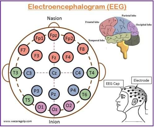
- About 16 and 25 electrodes are attached to the scalp of the person with a special paste or a cap with the electrodes is used.
- Once the recording begins, the person should remain still during the whole test. The technician will monitor the person from the window of an attached room to observe any movements such as swallowing or blinking that can cause an inaccurate reading.
- After the initial recording at rest, various stimuli are given to make brain wave activity that is not seen during rest. For example, asking the person to breathe deeply and rapidly or exposing to a bright flashing light.
- The test may take 45 to 120 minutes.
- The EEG is done during sleep if the evaluation is for a sleep disorder.
- For prolonged EEG monitoring the person may also be admitted to the hospital
- An ambulatory EEG is done when prolonged inpatient monitoring is not possible.
After the Procedure
- The electrodes are removed after the completion of the test and the electrode paste is washed off with warm water, acetone, witch hazel, or Seabreeze. A hair wash may also be done.
- The person who has been given sedatives may need rest until the effect has worn off and needed assistance to drive him/her home.
- Skin irritation or redness may be present at the placement of the electrodes were placed which wear off in a few hours.
- The person may resume any medicines that were held before taking the test.
Risks
- In rare cases, an EEG can cause seizures in a person with a seizure disorder due to the flashing lights or the deep breathing performed during the test.
- Certain factors or conditions may interfere with the interpretation of the test results. These include:
- Hypoglycemia (low blood sugar)
- Body movement or blinking during the test
- Bright or flashing lights
- Sedatives
- Caffeine-contained drinks like tea, coffee, or cola
- Oily hair, hair spray, or other hair products
Interpretation of the Result
The characteristics of EEG waveforms may be based on their frequency, location, morphology, amplitude, continuity, synchrony, symmetry, and reactivity. There are 4 types of waves in EEG according to the frequency that are Alpha, Beta, Delta, and Theta waves.
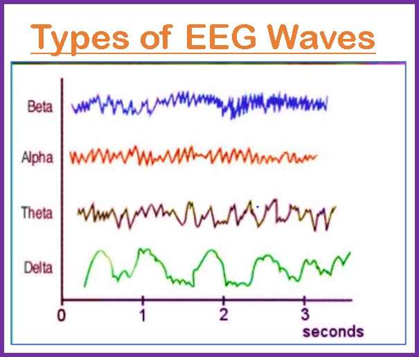
- Alpha waves occur in the occipital region and are found in a normal person who is in a resting but an awake stage. They indicate a state of calm and relaxation.
- Beta waves indicate an alert state such as when the person focuses on solving a problem. They are recorded from the frontal and parietal regions and are divided into 2 types:
- Beta 1 is constrained by cerebral activity.
- Beta 2 is excited by mental activity like tension.
- Theta waves are recorded in the temporal region and occur when the person is daydreaming and drowsiness. This indicates stress and frustration. Emotional questions are asked to get in these waves.
- Delta waves are recorded in the cortex region which occurs during deep sleep in premature babies and in cases of brain diseases. Occurs in persons who are in deep sleep or coma.
Normal results
- Electroencephalogram shows the electrical activity of our brain as a pattern of waves.
- Different levels of consciousness have a specific range of frequencies of waves per second which are considered normal.
- The wave patterns travel faster when the person is awake than when they are asleep.
- The EEG will reveal if the frequency patterns or waves are normal or not.
Abnormal results
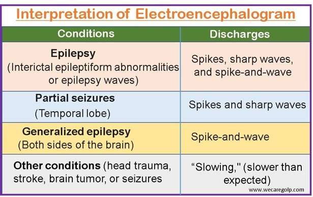
Abnormal EEG results may be due to:
- Epilepsy or any other seizure disorder
- Abnormal bleeding
- Sleep disorder
- Encephalitis
- Tumor
- Dead tissue due to a blockage of blood flow
- Migraine
- Excessive alcohol or drug use
- Head injury
Summary
- An EEG is a test/method that helps to detect abnormalities in the electrical activity or waves of the brain.
- During the procedure, electrodes are attached to the scalp which consists of small metal discs with thin wires to detect tiny electrical charges resulting from the activity of our brain cells.
- The process takes 30 to 60 minutes.
- The person’s hair should be free of oil, sprays, or any other hair products.
- The procedure does not have any significant risks.
- The test is performed in persons with a history of seizures.
References
- Casson, A.J., Abdulaal, M., Dulabh, M., Kohli, S., Krachunov, S., Trimble, E. (2018). Electroencephalogram. In: Tamura, T., Chen, W. (eds) Seamless Healthcare Monitoring. Springer, Cham. https://doi.org/10.1007/978-3-319-69362-0_2
- Craik, A., He, Y., Contreras-Vidal, J. L. (2019). Deep learning for electroencephalogram (EEG) classification tasks: a review. J. Neural Eng, 16(3), 1001. DOI 10.1088/1741-2552/ab0ab5
- WebMD. (n.d.). EEG (Electroencephalogram). Retrieved on 2023, Feb 23 from https://www.webmd.com/epilepsy/guide/electroencephalogram-eeg
- Blocka, K., Yetman. D. (2021, Nov 9). Electroencephalogram. Healthline. Retrieved on 2323, Feb 23 from https://www.healthline.com/health/eeg
- Britton, J.W., Frey, L.C., Hopp, J.L. (2016). Electroencephalography (EEG): An Introductory Text and Atlas of Normal and Abnormal Findings in Adults, Children, and Infants [Internet]. Chicago: American Epilepsy Society. https://www.ncbi.nlm.nih.gov/books/NBK390346/
- Sirven, J. I.. Schachter, S. C. (2013). Electroencephalography (EEG). Epilepsy Foundation. Retrieved on 2023, Feb 20 from https://www.epilepsy.com/diagnosis/eeg